Cell segmentation cellpose
Cell Segmentation: Cellpose
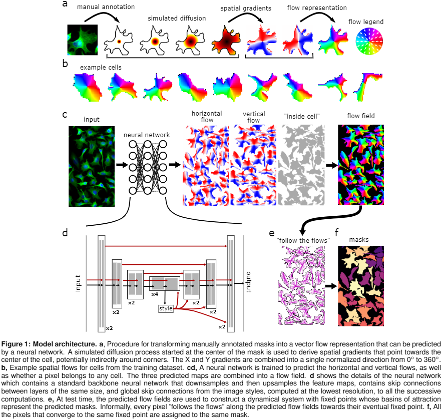
Cellpose is an anatomical segmentation algorithm for the identification of cells from high-resolution microscopy images, developed by Carsen Stringer and Marius Pachitariu.
Cellpose Parameters:
- Pretrained Model: Pretrained model to use for segmentation (e.g. cyto3)
- Channel Axis: Axis of image with color channels (e.g. 0 if image is (C, X, Y))
- Channel to Segment: Channel to segment
- Nuclear Channel: Channel to segment nuclei (optional for cyto models)
- Cell Diameter: If 0, will use the diameter of the training labels used in the model, or with built-in model will estimate diameter for each image
- Flow Error Threshold: 0 turns off this optional QC step, higher values will remove more cells
- Cell Probability Threshold: Decrease to find more and larger masks
- No Resample: Disable dynamics on full image (makes algorithm faster for images with large diameters)
- Exclude on Edges: Discard masks which touch edges of image
- Z Axis: Axis of image which corresponds to Z dimension (optional)
- Anisotropy of volume in 3D: Anisotropy of volume in 3D
Outputs:
measurements.csv: Spreadsheet with summary metrics for each of the detected cellscells.geo.json: Location of each detected cell in GeoJson format
Citations:
- Stringer C, Wang T, Michaelos M, Pachitariu M. Cellpose: a generalist algorithm for cellular segmentation. Nat Methods. 2021 Jan;18(1):100-106. doi: 10.1038/s41592-020-01018-x. Epub 2020 Dec 14. PMID: 33318659.
- Pachitariu M, Stringer C. Cellpose 2.0: how to train your own model. Nat Methods. 2022 Dec;19(12):1634-1641. doi: 10.1038/s41592-022-01663-4. Epub 2022 Nov 7. PMID: 36344832; PMCID: PMC9718665.
- Pachitariu M, Stringer C. Cellpose3: one-click image restoration for improved cellular segmentation. bioRxiv 2024.02.10.579780.
This pipeline supports viewing its output using the embedded Single-Cell Dashboard App directly on the output dataset's Overview page.